

Abstract
Gonadoblastoma is a rare gonadal tumor composed of sex cord cells and primitive germ cells. While the majority of gonadoblastomas are found in individuals with 46,XY gonadal dysgenesis, they are also rarely seen in patients with a 46,XX karyotype. We report a case of a 14.5 year-old girl presenting with an uncommon cause of virilization; a virilizing ovarian tumor. The patient underwent bilateral salpingo-oophorectomy. Upon histopathological examination, the excised tumor was confirmed to be bilateral gonadoblastoma, with dysgerminoma on the left side. Malignant gonadal tumors should be considered in cases of primary gonadal insufficiency with a 46,XX karyotype and progressive virilization. Even when laboratory and imaging tests show no abnormalities, a gonadal biopsy should be considered.
Keywords: Dysgerminoma, gonadoblastoma, virilization, XX gonadal dysgenesis
What is already known on this topic?
Gonadoblastomas typically affect individuals with 46,XY gonadal dysgenesis or females with Y chromosome material, rarely occurring in 46,XX women. Preventive gonadectomy is recommended for those with partial or complete gonadal dysgenesis due to the high malignancy risk associated with the Y chromosome material.
What this study adds?
The case expands the published evidence concerning gonadoblastoma by detailing its presentation, diagnosis, and management in a progressively virilized 46,XX patient. For progressive hirsutism and virilization without typical symptoms like abdominal pain, diagnostic laparoscopy and biopsy may be considered when conventional methods are inconclusive. Confirming the Y chromosomal material status is crucial when gonadal dysgenesis is suspected.
Introduction
Hyperandrogenism and the resulting manifestations in young female children can stem from various inherited and acquired causes. In some cases, androgen-producing tumors in the ovaries or adrenal glands can quickly lead to virilization. Gonadoblastomas are uncommon ovarian tumors composed of sex cord and primitive germ cell components. While gonadoblastomas are typically benign, they are often found alongside invasive germ cell malignancies. Typically, the tumor is observed in individuals with certain gonadal gene mutation syndromes, such as pure or mixed gonadal dysgenesis. The majority of gonadoblastomas are found in individuals with 46,XY gonadal dysgenesis, as well as in females carrying Y chromosome material. However, it is exceptionally rare for gonadoblastoma to occur in women with a normal 46,XX karyotype (, ). We present a case report of a 14.5-year-old girl with a normal 46,XX karyotype who was diagnosed with gonadoblastoma accompanied by dysgerminoma. We describe the clinical symptoms exhibited by the patient and the outcome of her treatment, along with a comprehensive analysis of previously reported cases of gonadoblastoma occurring in individuals with a typical 46,XX karyotype.
Case Report
A 14.5-year-old girl was referred to our outpatient clinic because of significant hirsutism developing over the preceeding two months. She was born at term, appropriate for gestational age, to first-degree consanguineous parents after an uneventful pregnancy, and her developmental milestones were normal. Puberty began at the age of 10.5 years, but menarche has not yet occurred.
At presentation, her weight, height, and body mass index (BMI) were 42.7 kg [-1.95 standard deviation score (SDS)], 158.1 cm (-0.5 SDS), and 17.08 kg/m2 (1.85 SDS), respectively. The pubertal stage was Tanner 4, and her external genitalia were remarkable for marked clitoromegaly, measuring 1.5x1 cm, with symmetrical labioscrotal folds and no palpable gonads. The modified Ferriman-Gallwey (mFG) score was 16, and a significant deepening of her voice was notable. Otherwise, the systemic examination was normal.
Hormonal evaluation revealed high serum luteinizing hormone (43.4 mIU/L) and follicle-stimulating hormone (87.6 mIU/L) levels, confirming the diagnosis of hypergonadotropic hypogonadism. In addition, serum total testosterone concentration was elevated (1.07 ng/mL). Basal levels of androgen precursors were normal (see ), and adrenal-derived hyperandrogenism was ruled out with a normal response to a corticotropin stimulation test. Tumor markers, including b-human chorionic gonadotropin (β-hCG), α-fetoprotein (αFP), and lactate dehydrogenase (LDH), were normal. Abdominal ultrasound (US) showed no abnormalities, and bilateral adrenal glands were normal. Pelvic US showed that the size of the uterus and ovaries were appropriate for the pubertal stage. In this hypergonadotropic hypogonadism patient with hyperandrogenism, cytogenetic analysis revealed a normal female karyotype (46,XX).
Table 1. Clinical, laboratory and radiological examination of the patient during follow up
- | At initial presentation | Before gonadectomy | After gonadectomy |
|---|---|---|---|
Age (years) | 14.6 | 16.3 | 17.2 |
Anthropometric evaluation | |||
Height, cm (SDS) | 158.1 (-0.5) | 159.5 (-0.51) | 159.5 (-0.6) |
Weight, kg (SDS) | 42.7 (-1.95) | 42.4 (-2.47) | 40.6 (-3.0) |
BMI, kg/m2 (SDS) | 17.08 (-1.85) | 16.6 (-2.72) | 15.9 (-3.7) |
Puberty Tanner Clitoromegaly | B4/4 P5 2-1.5 cm | B4/4 P5 3 cm | B5/5 P5 2.5x2 cm |
Ferriman-Gallwey score | 16 | 22 | NA |
Hormonal findings | |||
LH, mIU/mL (N=0.4-11.7) | 43.4 | 32 | 43.7 |
FSH, mIU/mL (N=1-9.2) | 87.6 | 85 | 86.6 |
E2, pg/mL (N=12.5-166) | 26.7 | 20 | 32 |
T, ng/mL (N=0.23-1.39) | 1.07 | 1.57 | 0.07 |
DHT, pg/mL (N=30-180) | 114 | NA | NA |
T/DHT | 9.3 | NA | NA |
AMH, ng/mL (N=0.62-7.8) | 3.05 | 3 | NA |
DHEA-S, ug/dL (N=44-248) | 331 | 317 | NA |
Cortisol, ug/dL (N=8-19) | 18.2 | NA | NA |
ACTH, pg/mL (N=6-48) | 19 | 17 | NA |
17-OHP, ng/mL (N=0.44-2.35) | 0.57 | NA | 0.8 |
1,4 AS, ng/mL (N=0.5-2.24) | 1 | 4.1 | 1.6 |
AS/T (N≥0.8) | 0.93 | 2.6 | 22.9 |
P, ng/mL (N=0.057-0.893) | 0.37 | NA | NA |
SHBG, nmol/L (N=36-125) | NA | 24.7 | NA |
CA 19.9, U/mL (N=0-34) | NA | NA | 13.6 |
β-hCG, mIU/mL (N≤5) | 0.93 | NA | NA |
AFP, ng/mL (N≤13.6) | 2.2 | 1.9 | 1.8 |
LDH, U/L (N=150-300) | 166 | 223 | 170 |
Imaging | |||
Pelvic US | Uterus: 41x17x65 mm Right adnexa: 9.5 mL Left adnexa: 10.9 mL | Uterus: 28x13.3x63 mm cervix/fundus:1 Adnexa: Bilateral ovaries could not be clearly distinguished. Isoechoic tissue areas containing cystic openings, which may be compatible with the follicle cyst, whose volume was measured as 1.1 mL on the right and 1 ml on the left, were observed in the ovarian lobes. Fibrotic ovarian tissue? | - |
Pelvic MRI | Uterus: Uterine size, parenchyma and echogenicity are normal Ovaries: Could not be seen | Uterus: 43x18x11 mm, its dimensions are reduced. Ovaries: Bilateral ovaries were not seen. | |
Histopathology | |||
- | - | Bilateral gonadoblastoma and unilateral (left) dysgerminoma | - |
ACTH: Adrenocorticotropic hormone, AFP: α-fetoprotein, AMH: Anti-Mullerian hormone, BMI: Body mass index, CA 19-9: Carbohydrate antigen 19-9, 1,4 AS: 1,4 Androstenedione, DHEA-S: Dehydroepiandrosterone sulfate, DHT: Dihydrotestosterone, E2: Estradiole, FSH: Follicle-stimulating hormone , β-hCG: Human chorionic gonadotrophin, LDH: Lactate dehyrogenase, LH: Luteinizan hormone, 17-OHP: 17-hydroxy progesterone, N: Normal value, NA: Not avaliable, P: Progesterone, SHBG: Sex hormone binding globuline, T: Total testosterone, US: Ultrasonography, MRI: Magnetic resonance imaging
Unfortunately, the patient missed follow-up visits for two years. At the age of 16.3 years, she was re-evaluated. During the past two years, she had become increasingly uncomfortable due to progressive hirsutism and needed laser hair removal session every two weeks on her whole body. At this time, the mFG score was 22, and clitoral length was measured at 3 cm. Fibrotic ovarian tissue was detected; however, no abdominal or gonadal mass was visualised on US. Abdominal magnetic resonance imaging (MRI) revealed similar findings (as shown in ). Fluorescence in situ hybridization analysis showed no alterations in the SRY gene. Polymerase chain reaction (PCR) analysis for the SRY gene sequence in the DNA sample obtained from ovarian tissue did not show the presence of SRY. In addition, whole exome sequencing was performed to comprehensively assess all known genetic disorders, with a particular focus on sex-determining genes, such as WT1, SOX9, and DAX1. A laparoscopic gonad biopsy was performed and it revealed bilateral gonadoblastoma and unilateral (left) dysgerminoma. Ultimately, the patient underwent bilateral oophorectomy. Macroscopic pathological examination of the salpingo-oophorectomy material revealed a right ovary measuring 3×1×0.5 cm, while the left ovary was slightly larger, measuring 2.7×1.5×3 cm.
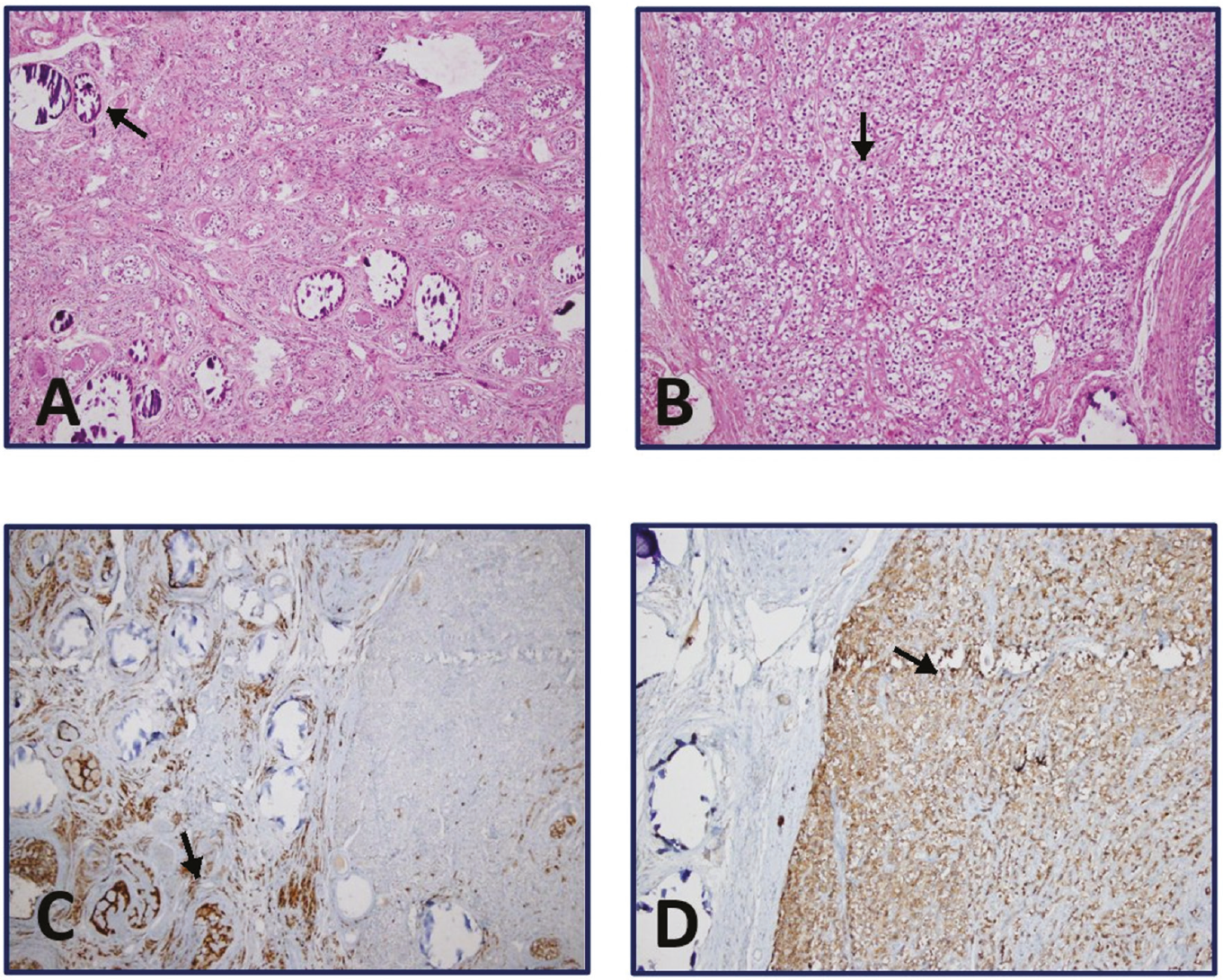
Histopathological microscopic analysis provided further insight into the cellular composition. The specimen revealed the presence of primitive germ cells alongside sex cord stromal cells, surrounded by ovarian-type stroma. Notably, morphological analysis identified a dysgerminoma in the left ovary. This tumor exhibited characteristic cell cords, featuring both clear and eosinophilic cytoplasm, and measured approximately 0.5 cm in diameter. Immunohistochemical staining was performed to further classify the cellular components. The sex cord cells within the gonadoblastoma stained positively for inhibin, confirming their identity. In contrast, the germ cells within the dysgerminoma stained positively for C-kit, which is a known marker for germ cell tumors. These staining patterns facilitated the differentiation of the various cell types present within the tumor (see ). No pathological F-18 fluorodeoxyglucose uptake was detected on positron emission tomography/computed tomography, and she did not require or receive any chemotherapy. The bone mineral density z-score was -1.7, and hormone replacement therapy was commenced following gonadectomy. Pelvic MRI and tumor markers were subsequently checked at 6-month intervals, with no abnormal findings evident.
Discussion
We describe a girl with a 46,XX karyotype who exhibited virilization, a condition rarely caused by gonadoblastoma. The etiology of virilization in this girl was clinically challenging due to the variety of potential causes. Various inherited and acquired conditions may contribute to virilization, including disorders of sex development (DSD), virilizing ovarian tumors, adrenal tumors, and exposure to exogenous androgens. Among the DSD conditions, different forms of congenital adrenal hyperplasia, ovotesticular DSD, and aromatase deficiency may manifest with hyperandrogenic features in females. These conditions contribute to the clinical spectrum of causes that need to be considered when evaluating virilization in a young girl. The severity of virilization served as an important clinical indicator, suggesting an ovarian cause as the underlying factor in this girl. The rapid and severe nature of the virilization, combined with the exclusion of adrenal causes by a normal corticotropin stimulation test and normal adrenal precursor levels, directed the clinical suspicion toward an androgen-secreting ovarian tumor.
Gonadoblastoma was first recognized as a distinct entity by Scully () in 1953. Histologically, it is characterized by the presence of distinct clusters consisting of germ cells and sex cord elements that resemble immature Sertoli or granulosa cells. Histopathologically, the present case exhibited sex cord cells in the gonadoblastoma and germ cells in the dysgerminoma.
Gonadoblastoma has a high incidence in patients with various syndromes of gonadal maldevelopment that are characterized by the presence of the Y chromosome (). Gonadoblastomas are observed in approximately 25% to 30% of patients with XY gonadal dysgenesis and in approximately 15% to 20% of individuals with a karyotype of 45,X/46,XY (). However, a number of cases have been reported in females with a normal 46,XX karyotype. Pratt-Thomas and Cooper () reported a case where unilateral gonadoblastoma that was found in association with a ruptured ectopic tubal pregnancy, and the cytogenetic evaluation of peripheral leukocytes revealed a karyotype of 46,XX/45,XO.
In individuals with a 46,XX karyotype, the mechanism underlying the development of gonadoblastoma in the absence of a Y chromosome remains unclear. However, potential contributing factors may include somatic mutations, epigenetic changes, or hormonal influences. Somatic mutations in genes such as WT1 and SF1 may disrupt gonadal development, leading to tumorigenesis. Epigenetic alterations, including DNA methylation or histone modifications, may silence or activate genes inappropriately, leading to abnormal gonadal development (, ). Hormonal influences, such as elevated gonadotropin levels, are thought to potentially play a role in the development of gonadoblastoma but the precise mechanisms underlying this association have yet to be fully elucidated (). Future studies are essential to elucidate these alternative pathways, and understanding such mechanisms may reveal novel targets for early detection and treatment in the 46,XX population of patients with gonadoblastoma.
While gonadoblastomas are typically considered benign, they often coexist with invasive germ cell malignant tumors. The most prevalent malignancy found in association with gonadoblastomas is pure dysgerminoma. However, other variants, such as immature teratoma, embryonal carcinoma, yolk sac tumor, and choriocarcinoma, have also been reported. In Scully’s review of 74 gonadoblastoma cases approximately 50% of the tumors were accompanied by dysgerminoma as a coexisting germ cell tumor. This is similar to the presented case and consistent with previous studies. Other types of germ cell tumors, such as embryonal carcinoma, yolk sac tumor, and teratoma, were also reported in a few cases (, ).
In our review of the literature since 1990, we identified 14 cases of gonadoblastoma in females with a normal 46,XX karyotype (,,,,,,,,,,,,,). presents a summary of the clinical characteristics observed in these 14 cases as documented in the literature and also includes the presented case for comparison. While abdominal symptoms, such as abdominal pain or the presence of a mass, were the most common complaints in the 15 reported cases (66%, 10/15 cases), it is important to note that cases have also presented with symptoms such as vaginal bleeding, menstrual disorders, and virilization. Our case unusual because of several distinctive findings. Firstly, there was an absence of abdominal pain, which is a common complaint in similar cases. Moreover, both physical examination and imaging tests (US and MRI) did not identify any masses. Furthermore, the levels of tumor markers, including αFP, β-HCG, and LDH were not significantly elevated. Given these unique characteristics, an important aspect of the present case was that the diagnosis made through diagnostic laparoscopy. In the case reported by Chandrapattan et al. (), and similar to our case, the chief complaint was virilization. Furthermore, contrasexual pubertal development was observed in that particular case.
Table 2. A review of published female cases of gonadoblastoma with karyotype 46,XX
No | Author | Year | Age at diagnosis | Clinical presentation | Fertile status | Hormonal abnormalities | Laterality | Karyotype | Tumor size | Surgical procedure | Pathological findings in addition to gonadoblastoma | Adjuvant therapy |
|---|---|---|---|---|---|---|---|---|---|---|---|---|
1 | Erhan et al. () | 1992 | 26 | Abdominal mass during pregnancy | Fertile | NR | Right | 46, XX | 20 cm | TAH + BSO | Dysgerminoma | Chemotherapy |
2 | Obata et al. () | 1995 | 10 | Abdominal discomfort | No | NR | Bilateral | 46, XX | 8 cm | USO + right Ovarian cystectomy | Left with dysgerminoma, right with dysgerminoma and yolk sac tumor | Chemotherapy |
3 | Zhao et al. () | 2000 | 27 | Abdominal pain | Fertile | NR | Right | 46, XX | 10x8x6 cm | USO + chemotherapy + later TAH + USO + LND + omentectomy | Choriocarcinoma, embryonal carcinoma, yolk sac tumor, immature teratoma and dysgerminoma | Chemotherapy |
4 | Erdemoglu and Ozen () | 2007 | 19 | Abdominal mass and pain | No | NR | Unilateral | 46, XX | 25x22x20 cm | USO | Endodermal sinus tumor | None |
5 | Yilmaz et al. () | 2010 | 20 | Abdominal mass | No | NR | Bilateral | 46, XX | 21x21x11 cm | BSO | Bilateral with dysgerminoma | Radiation and chemotherapy |
6 | Koo et al. () | 2011 | 34 | Vaginal bleeding | Fertile | NR | Left | 46, XX | 12 x9x 7 cm | USO + paraaortic LND | Dysgerminoma | Chemotherapy |
7 | Esin et al. () | 2011 | 15 | Vaginal bleeding | No | NR | Left | 46, XX | 0.5 cm | USO | Dysgerminoma | None |
8 | Kanagal et al. () | 2013 | 14 | Abdominal distension | No | Elevated testosterone level | Left | 46, XX | 30x25x10 cm | USO + pelvic and para-aortic LND + infracolic omentectomy | Dysgerminoma | Chemotherapy |
9 | Kulkarni et al. () | 2016 | 20 | Abdominal pain | Fertile | NR | Left | 46, XX | 8x6x6 cm | USO + omental biopsy | Dysgerminoma | None |
10 | McCuaig et al. () | 2017 | 20 | Oligomenorrhea and menorrhagia | No | NR | Left | 46, XX | 2.5x2x1.5 cm | USO | Dysgerminoma | None |
11 | Roth et al. () | 2019 | 9 | Abdominal pain and a right adnexal mass | No | NR | Right | 46, XX | 8.9×5.7×5.2 cm | USO | Mixed germ cell tumor | Chemotherapy |
12 | Raafey et al. () | 2020 | 10 | Abdominal pain, abdominal distention and fever | No | NR | Left | 46, XX | 16x10x9 cm | USO | Dysgerminoma | Chemotherapy |
13 | Chandrapattan et al. () | 2022 | 9 | Signs of virilization and contrasexual pubertal development | No | NR | Right | 46, XX | 15x10 cm | USO + pelvic LND + infracolic omentectomy | Dysgerminoma | Chemotherapy |
14 | Yin et al. () | 2022 | 11 | Abdominal pain | NR | Elevated FSH, LH, progesterone, 11-deoxycortisole, ACTH, corticosterone decreased cortisol, E2, testosterone | Right | 46, XX, gonadal karyotype Y chromosome negative | 10x8x6 cm | USO | Dysgerminoma | 17a-hydroxylase /17,20-lyase deficiency Glucocorticoid replacement therapy |
15 | Present case | 2024 | 14.6 | Hirsutism | No | Elevated FSH, LH, total testosterone | Bilateral | 46, XX SRY negative | 3x1x0.5 cm (right) 2.7x1.5x3 cm (left) 0.5 cm (left) dysgerminoma | BSO | Bilateral gonadoblastoma and unilateral (left) dysgerminoma | None |
BSO, bilateral salpingo-oophorectomy; USO, unilateral salpingo-oophorectomy; LND, lymph node dissection; TAH, total abdominal hysterectomy; NR, not report
In individuals with partial or complete gonadal dysgenesis, preventive bilateral gonadectomy is commonly recommended due to the markedly increased risk of gonadal malignancy (15% to 50%) associated with the presence of the Y chromosome (). Therefore, it is important to confirm if Y chromosomal material is present or not. Typically, peripheral blood lymphocyte karyotyping is performed to detect Y chromosomes in the germline but this approach carries the risk of missing small chromosomal fragments containing Y chromosome material. In the presented case, PCR analysis for the SRY gene sequence in the DNA sample obtained from the ovarian tissue did not demonstrate the presence of SRY.
Surgery is the primary treatment method for the management of gonadoblastomas. The specific surgical approach may vary depending on the extent and characteristics of the tumor. It is important to consider individual patient factors and provide appropriate adjunct therapies, such as chemotherapy, to support the overall treatment plan. Out of the 14 previously documented cases, chemotherapy was administered in nine (64.3%) cases. Due to the localized and small nature of the tumor in our patient, chemotherapy was deemed unnecessary.
Conclusion
It is important to consider that when managing hirsutism and progressive virilization, laparoscopy should be performed so that biopsies may provide guidance when imaging methods and laboratory tests fail to yield a definitive diagnosis. Further studies are needed to elucidate the etiopathogenesis of reported cases.
Footnotes
Ethics
Informed Consent: Informed consent for publication was obtained from the patient’s parents.
Authorship Contributions
Surgical and Medical Practices: Ozlem Dural, Ayca Dilruba Aslanger, Aysel Bayram, Semen Onder, Concept: Tugce Kandemir, Design: Tugce Kandemir, Data Collection or Processing: Tugce Kandemir, Esin Karakilic Ozturan, Analysis or Interpretation: Tugce Kandemir, Literature Search: Tugce Kandemir, Writing: Tugce Kandemir, Esin Karakilic Ozturan, Elif Inan Balci, Asli Derya Kardelen, Melek Yildiz, Sukran Poyrazoglu, Firdevs Bas, Feyza Darendeliler.
Conflict of Interest
Conflict of Interest: One author of this article, Feyza Darendeliler, is a member of the Editorial Board of the Journal of Clinical Research in Pediatric Endocrinology. However, she did not involved in any stage of the editorial decision of the manuscript. The editors who evaluated this manuscript are from different institutions. The other authors declared no conflict of interest.
Financial Disclosure
Financial Disclosure: The authors declared that this study received no financial support.
References
Scully RE. Gonadoblastoma; a gonadal tumor related to the dysgerminoma (seminoma) and capable of sex-hormone production. Cancer. 1953;6:455-463.
Kim M-J, Ahn H-J, Kim J-Y, Kim K-R. Gonadoblastoma overgrown by dysgerminoma in women with 46, XX karyotype. The Korean Journal of Pathology. 2003;37:66-70.
Pratt-Thomas HR, Cooper JM. Gonadoblastoma with tubal pregnancy. Am J Clin Pathol. 1976;65:121-125.
Ferrari MTM, Silva ESDN, Nishi MY, Batista RL, Mendonca BB, Domenice S. Testicular differentiation in 46,XX DSD: an overview of genetic causes. Front Endocrinol (Lausanne). 2024;15:1385901.
Costanzo M, Touzon MS, Marino R, Guercio G, Ramirez P, Mattone MC, Pérez Garrido N, Bailez MM, Vaiani E, Ciaccio M, Galluzzo Mutti ML, Belgorosky A, Berensztein E. Gonadal tumor development in 46,XX disorders of gonadal development. Eur J Endocrinol. 2022;187:451-462.
Looijenga LH, Hersmus R, Oosterhuis JW, Cools M, Drop SL, Wolffenbuttel KP. Tumor risk in disorders of sex development (DSD). Best Pract Res Clin Endocrinol Metab. 2007;21:480-495.
Obata NH, Nakashima N, Kawai M, Kikkawa F, Mamba S, Tomoda Y. Gonadoblastoma with dysgerminoma in one ovary and gonadoblastoma with dysgerminoma and yolk sac tumor in the contralateral ovary in a girl with 46XX karyotype. Gynecol Oncol. 1995;58:124-128.
Erhan Y, Toprak AS, Ozdemir N, Tiras B. Gonadoblastoma and fertility. J Clin Pathol. 1992;45:828-829.
Zhao S, Kato N, Endoh Y, Jin Z, Ajioka Y, Motoyama T. Ovarian gonadoblastoma with mixed germ cell tumor in a woman with 46, XX karyotype and successful pregnancies. Pathol Int. 2000;50:332-335.
Yilmaz B, Gungor T, Bayramoglu H, Soysal S, Mollamahmutoglu L. Bilateral ovarian gonadoblastoma with coexisting dysgerminoma in a girl with 46, XX karyotype. J Obstet Gynaecol Res. 2010;36:697-700.
Koo YJ, Chun YK, Kwon YS, Lee IH, Kim TJ, Lee KH, Lim KT. Ovarian gonadoblastoma with dysgerminoma in a woman with 46XX karyotype. Pathol Int. 2011;61:171-173.
Erdemoglu E, Ozen S. Ovarian gonodoblastoma with yolk sac tumor in a young 46, XX female: case report. Eur J Gynaecol Oncol. 2007;28:516-518.
Esin S, Baser E, Kucukozkan T, Magden HA. Ovarian gonadoblastoma with dysgerminoma in a 15-year-old girl with 46, XX karyotype: case report and review of the literature. Arch Gynecol Obstet. 2012;285:447-451. Epub 2011 Aug 31
Kanagal DV, Prasad K, Rajesh A, Kumar RG, Cherian S, Shetty H, Shetty PK. Ovarian Gonadoblastoma with Dysgerminoma in a Young Girl with 46, XX Karyotype: A Case Report. J Clin Diagn Res. 2013;7:2021-2022. Epub 2013 Aug 10
Kulkarni MM, Sinai Khandeparkar SG, Joshi AR, Bhayekar PV. Unilateral gonadoblastoma with dysgerminoma in normal fertile woman having a child: Extremely rare occurrence with characteristic immunohistomorphology. Indian J Pathol Microbiol. 2016;59:527-529.
McCuaig JM, Noor A, Rosen B, Casper RF, Mitri F, Colgan T, Kim RH. Case Report: Use of tumor and germline Y chromosomal analysis to guide surgical management in a 46, XX female presenting with gonadoblastoma with dysgerminoma. Int J Gynecol Pathol. 2017;36:466-470.
Roth LM, Davis MM, Czernobilsky B. Classic and “dissecting” gonadoblastoma in a phenotypic girl with a 46, XX peripheral karyotype and no evidence of a disorder of sex development. Int J Gynecol Pathol. 2019;38:581-587.
Raafey MA, Abdulwaasey M, Fatima SS, Uddin Z, Tariq MU. Bilateral gonadoblastoma with dysgerminoma in a phenotypically normal female with 46XX karyotype: report of a rare case and literature review. Cureus. 2020;12:e8990.
Chandrapattan P, Jena A, Patnayak R, Mangaraj S, Naik S, Panda S. Gonadoblastoma with dysgerminoma presenting as virilizing disorder in a young child with 46, XX karyotype: a case report and review of the literature. Case Rep Endocrinol. 2022;2022:5666957.
Yin M, Yang J, Tian Q, Zhang X. Ovarian gonadoblastoma with dysgerminoma in a girl with 46,XX karyotype 17a-hydroxylase/17, 20-lyase deficiency: A case report and literature review. Front Endocrinol (Lausanne). 2022;13:989695.