

Abstract
Objective
The hypothalamic-pituitary-gonadal axis is active during minipuberty, the timing of which coincides with infantile colic. To the best of our knowledge, the relationship between these entities has not been previously investigated.
Methods
Saliva samples were collected from 15- to 60-day-old term infants (n=139) between 9 am and 5 pm. Group 1 included infants with infantile colic (n=68, 54.4% female) while the remaining healthy infants constituted Group 2 (n=71, 47.9% female). Salivary levels of estradiol (Esal) in females and testosterone (Tsal) in males were measured by ELISA in duplicate.
Results
The median (25th-75th centile) age and birth week for all infants were 33 (29-43) days and 39 (38.1-40) weeks, respectively. Levels of Tsal in males [Group 1, 73.35 (59.94-117.82) pg/mL vs Group 2, 77.66 (56.49-110.08) pg/mL, p=0.956] and Esal in females [Group 1, 3.91 (2.76-5.31) pg/mL vs Group 2, 4.03 (1.63-12.1) pg/mL, p=0.683] were similar. However, in subjects with infantile colic (Group 1), Esal and body mass index (BMI) standard deviation scores of females were slightly correlated (Group 1, rs= 0.393, p=0.016 vs. Group 2, rs= 0.308, p=0.076) and there was a significant correlation between the sampling time and Tsal in males (Group 1, rs= 0.469, p=0.009 vs. Group 2, rs= -0.005, p=0.976).
Conclusion
Random salivary sex steroid levels were similar in infants with and without infantile colic. However, in subjects with infantile colic, Esal levels in females were positively correlated with BMI and Tsal levels were higher later in the day among males. Thus, sex steroid production may be altered during minipuberty in subjects with infantile colic.
Keywords: Gonadal activity, newborn, puberty, fussing
What is already known on this topic?
The timings of minipuberty and infantile colic may coincide. There exists no data regarding the relationship between them.
What this study adds?
Sex steroid production may be altered during minipuberty in subjects with infantile colic.
Introduction
Classical puberty begins in adolescence. However, the hypothalamic-pituitary-gonadal axis is also active during the first months of life resulting in an increase in gonadotropins and sex steroids (minipuberty) (, , , ). In boys, the increase in follicle stimulating hormone (FSH) and luteinizing hormone (LH), primarily LH, reaches its highest value at 4-10 weeks after birth and both decrease to prepubertal levels at around six months. Serum testosterone level, which increases with LH secretion, peaks between 1-3 months and decreases to prepubertal levels between 6-9 months (, ). Similarly in girls, serum estradiol levels peak between 30-60 days of life, then fall below the prepubertal level towards one year of age (). In the literature, there are data regarding salivary testosterone levels in male infants, but data on salivary estradiol levels in female infants are limited (, , , ). The importance of minipuberty is not fully understood but minipuberty has been reported to affect genital organ development, body composition, and cognitive functions ().
Infantile colic was defined by Wessel et al. () in 1954 as excessive irritability and crying in the evening for no apparent reason, starting in the first weeks of life. It is seen in 10-40% of otherwise healthy infants aged one to five months. While various hypotheses, including gastrointestinal, neurodevelopmental, and psychosocial causes have been proposed, its etiology is currently unknown (, ).
Among the few and heterogeneous data regarding the impact of minipuberty on behavior, there are some findings indicating that sex steroid levels might be associated with behavioral patterns in infants as well as in adolescents (, , , ). Since sex steroid levels are elevated during a period when infantile colic is common, the aim was to evaluate the relationship between minipuberty and infantile colic by measuring testosterone and estradiol levels in saliva samples of infants. To the best of our knowledge, there is no similar published study.
Methods
Subjects
This study included term infants aged 15-60 days old without any additional disease who attended outpatient clinics in a single center between March and October 2021. Infants who had any disorder of sex development or a systemic pathology, such as gastrointestinal malformation and gastroesophageal reflux, or who had used antibiotics in the last week were not included in the study. Infantile colic was diagnosed according to the Rome IV criteria (episodes of crying and irritability lasted longer than one week, at least three hours a day, and at least three days in the same week) (, ). Careful physical examinations of these infants were performed to exclude other systematic causes of irritability. All of the infants were prepubertal and none had an abnormal external genital structure. The parents of the subjects with infantile colic were contacted by phone when they were six months old and it was confirmed that they did not have any other diseases.
Data Collection
All relevant data, including demographic features, family history, anthropometric measurements, and physical examination findings were recorded. Standard deviation scores (SDS) for weight, length, head circumference, and body mass index (BMI) were calculated according to the Turkish child population using child metrics (, ). Weight for length SDS was calculated according to World Health Organization data (, ).
Saliva samples were collected using Salimetrics® SalivaBio Oral Swab (Salimetrics, State College, PA, USA) and stored at -80 °C. Measurement was made with Salimetrics® 17 b-estradiol ELISA kit for estradiol (1-3701) in girls and Salimetrics® Testosterone ELISA kit for testosterone (1-2402) in boys and all samples were tested in duplicate. When the first and second results were statistically compared for testosterone and estradiol, p values were 0.922 and 0.347, respectively. An average of the two measurements were used in the study. Both kits are based on a sandwich ELISA method and are read at a wavelength of 450 nm. The sensitivity of the estradiol kit was 0.1 pg/mL, and the testosterone kit was 1 pg/mL. The measurement ranges were 1-32 pg/mL for estradiol and 6.1-600 pg/mL for testosterone.
Ethics
This study was conducted with the approval of Dokuz Eylül University Local Ethical Committee (decision no: 2019/22-22, date: 09.09.2019). Financial support was provided by the Department of Scientific Research Projects of Dokuz Eylül University (2020.KB.SAG.40) and Turkish Society for Pediatric Endocrinology and Diabetes (2020-04). An informed written consent form was obtained from parents before participating the study and it was performed in accordance with the principles of the Declaration of Helsinki.
Statistical Analysis
In order to be able to find a significant difference of 5 pg/mL between the mean of sex steroids of the groups in a situation where the standard deviation values of the groups were 4 and 8 pg/mL, based on a type 1 error of 0.05 and a power of 0.80, the minimum number of subjects for each gender among the groups was determined as 27. Taking unexpected errors into account, it was planned to include 20% excess cases.
Statistical analysis was performed using IBM Statistical Package for the Social Sciences, version 24 (IBM Inc., Armonk, NY, USA). The distribution of the data was evaluated using the Kolmogorov-Smirnov test. Descriptive statistics are given as numbers and percentages for categorical variables and median (25th-75th centile) for numerical variables. The Mann-Whitney U test was used to compare numeric variables and chi-square test was used for categorical data. The correlation of the parameters was tested with Spearman correlation analysis. A p value <0.05 was considered significant.
Results
A total of 139 infants (48.9% males) were included in the study. The median age was 33 (29-43) days, and the median gestational age was 39 (38.1-40) weeks. A majority of the newborns (n=88, 63.3%) were born by caesarean section.
In terms of feeding, 100 (71.9%) were fed with breast milk only, 35 (25.2%) with both breast milk and formula, and four (2.9%) with formula only. The median value of daily stool counts was 3 (2-4; 1-9). Only three (2.2%) were not using any medication, 120 (86.3%) were given vitamin D, 15 (10.8%) were given vitamin D and probiotics and one (0.7%) was only using probiotics.
The subjects were divided into two groups: with infantile colic (Group 1, n=68); and those without (Group 2, n=71). The infants in the two groups had similar demographic and anthropometric features, except for gestational age and weight-for-length SDS (). In addition, the ages, education, health status, drug use, and smoking rates of parents were similar between the groups (data not shown).
Table 1. The demographic and anthropometric characteristics regarding presence of infantile colic. Group 1: subjects with infantile colic, Group 2: subjects without infantile colic
Group 1 (n=68) | Group 2 (n=71) | p | |
|---|---|---|---|
Age (in days) | 34.5 (29-43.8) | 33 (29-43) | 0.439 |
Gender n (%) Female Male | 37 (54.4) 31 (45.6) | 34 (47.9) 37 (52.1) | 0.442 |
Mode of delivery n (%) SVD C/S | 21 (30.9) 47 (69.1) | 30 (42.3) 41 (57.7) | 0.164 |
Gestational age | 38.5 (38-39.6) | 39.2 (38.5-40.2) | <0.001 |
Birth weight SDS | -0.03 [(-0.43)-0.61] | 0.01 [(-0.72)-0.52] | 0.903 |
Birth length SDS | 0.00 [(-0.38)-0.75] | -0.19 [(-0.66)-0.45] | 0.089 |
Birth head circumference SDS | 0.07 [(-0.36)-0.43] | 0.07 [(-0.64)-0.79] | 0.899 |
Weight SDS | 0.4 [(-0.2)-0.84] | 0.15 [(-0.32)-0.62] | 0.168 |
Length SDS | 0.16 [(-0.48)-0.77] | 0.23 [(-0.34)-0.91] | 0.634 |
BMI SDS | 0.13 [(-0.11)-0.75] | 0.03 [(-0.67)-0.52] | 0.056 |
Weight for length SDS | 0.3 [(-0.16)-1.11] | -0.04 [(-0.87)-0.79] | 0.021 |
Head circumference SDS | 0.04 [(-0.52)-0.68] | -0.06 [(-0.71)-0.60] | 0.607 |
Data are presented as median (25p-75p) unless otherwise indicated.
SVD: spontaneous vaginal delivery, C/S: cesarean section, SDS: standard deviation score, BMI: body mass index
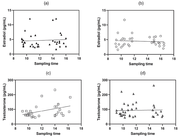
Characteristics of the subjects were further analyzed according to gender. Although it was observed that salivary estradiol levels decreased with increasing age in female babies (n=71), this did not reach statistical significance (rs=-0.224, p=0.061) (). Females with infantile colic (Group 1-F) were born slightly earlier compared to healthy control female subjects (Group 2-F), while the remaining features, including salivary estradiol levels, were similar (). Correlation analyses in females revealed that saliva estradiol levels showed significant correlation with BMI SDS in Group 1 only ().
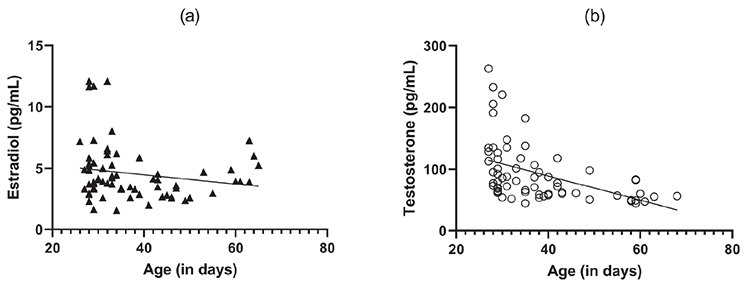
Table 2. The demographic and anthropometric characteristics regarding presence of infantile colic in females. Group 1-F: female subjects with infantile colic, Group 2-F: females without infantile colic
Group 1-F (n=37) | Group 2-F (n=34) | p | |
|---|---|---|---|
Age (in days) | 32 (29-43) | 34 (29-47) | 0.522 |
Mode of delivery n (%) SVD C/S | 11 (29.7) 26 (70.3) | 14 (41.2) 20 (58.8) | 0.313 |
Gestational age | 38.28 (37.71-39.21) | 39 (38.42-39.88) | <0.001 |
Birth weight SDS | -0.14 [(-0.62)-0.46] | -0.11 [(-0.72)-0.52] | 0.773 |
Birth length SDS | -0.19 [(-0.42)-0.75] | -0.19 [(-0.66)-0.75] | 0.619 |
Birth head circumference SDS | 0.36 [(-0.36)-0.36] | -0.18 [(-0.36)-0.36] | 0.743 |
Crying frequency (day/week) | 5 (4-6) | 0 (0-5) | <0.001 |
Last feeding time (min) | 30 (17.5-60) | 30 (20-60) | 0.902 |
Sampling time n (%) 9:00-12:00 13:00-17:00 | 21 (56.8) 16 (43.2) | 19 (55.9) 15 (44.1) | 0.941 |
Weight SDS | 0.41 [(-0.23)-0.82] | 0.12 [(-0.29)-0.77] | 0.542 |
Length SDS | 0.02 [(-0.5)-0.76] | 0.26 [(-0.71)-0.91] | 0.936 |
BMI SDS | 0.13 [(-0.09)-0.77] | 0.36 [(-0.55)-0.70] | 0.881 |
Weight for length SDS | 0.32 [(-0.15)-1.18] | 0.06 [(-0.82)-1.02] | 0.208 |
Head circumference SDS | -0.08 [(-0.78)-0.69] | -0.19 [(-0.72)-0.62] | 0.995 |
Weight gain (g/day) | 33.75 (27.64-38.44) | 32.61 (23.75-40.13) | 0.782 |
Salivary estradiol (pg/mL) | 3.91 (2.76-5.31) | 4.03 (3.22-5.40) | 0.683 |
Data are presented as median (25p-75p) unless otherwise indicated.
SVD: spontaneous vaginal delivery, C/S: cesarean section, SDS: standard deviation score, BMI: body mass index
Table 3. Correlation of salivary estradiol level with demographic and clinical parameters in females, Group 1-F: females with infantile colic, Group 2-F: females without infantile colic
All females (n=71) | Group 1-F (n=37) | Group 2-F (n=34) | |
|---|---|---|---|
Age (in days) | rs=-0.224 (p=0.061) | rs=-0.142 (p=0.4) | rs=-0.268 (p=0.125) |
Birth length SDS | rs=-0.11 (p=0.361) | rs=-0.007 (p=0.965) | rs=-0.211 (p=0.231) |
Sampling time | rs=0.017 (p=0.887) | rs=-0.003 (p=0.987) | rs=0.042 (p=0.811) |
Weight SDS | rs=0.132 (p=0.274) | rs=0.109 (p=0.521) | rs=0.181 (p=0.305) |
BMI SDS | rs=0.346 (p=0.003) | rs=0.393 (p=0.016) | rs=0.308 (p=0.076) |
Weight for length SDS | rs=0.241 (p=0.043) | rs=0.287 (p=0.085) | rs=0.215 (p=0.221) |
Crying frequency (day/week) | rs=-0.078 (p=0.518) | rs=-0.216 (p=0.2) | rs=-0.009 (p=0.962) |
SDS: standard deviation score, BMI: body mass index
In male infants, salivary testosterone levels decreased with increasing age (). BMI SDS and weight-for-length SDS were found to be significantly higher in the males with infantile colic (Group 1-M); although salivary testosterone levels were similar between the groups (). Correlation analyses were also done for male babies (). There was a moderate negative correlation between testosterone and BMI SDS but only in the infantile colic group. When the correlation analysis of the sample collection time and testosterone level was examined, it was found that subjects in the infantile colic group whose samples were taken later in the day had higher salivary testosterone levels (). There was no similar association in the control group. In addition, when examined with partial correlation by controlling for age, the relationship between testosterone and sample collection time was found to be stronger (rs=0.469, p=0.009).
Table 4. The demographic and anthropometric characteristics regarding presence of infantile colic in males. Group 1-M: male subjects with infantile colic, Group 2-M: males without infantile colic
Group 1-M (n=31) | Group 2-M (n=37) | p | |
|---|---|---|---|
Age (in days) | 37 (31-49) | 31 (29-39.5) | 0.086 |
Mode of delivery n (%) SVD C/S | 10 (32.3) 21 (67.7) | 16 (43.2) 21 (56.8) | 0.353 |
Gestational age | 39 (38.28-40) | 39.71 (38.7-40.4) | 0.139 |
Birth weight SDS | 0.13 [(-0.25)-0.76] | 0.09 [(-0.66)-0.50] | 0.542 |
Birth length SDS | 0.00 [(-0.45)-0.91] | 0.00 [(-0.91)-0.45] | 0.072 |
Birth head circumference SDS | 0.07 [(-0.29)-0.79] | 0.07 [(-0.64)-0.79] | 0.895 |
Crying frequency (day/week) | 5 (4-6) | 0 (0-4.5) | <0.001 |
Last feeding time (min) | 60 (30-90) | 60 (30-105) | 0.369 |
Sampling time n (%) 9:00-12:00 13:00-17:00 | 16 (51.6) 15 (48.4) | 26 (70.3) 11 (29.7) | 0.115 |
Weight SDS | 0.38 [(-0.10)-0.86] | 0.16 [(-0.36)-0.55] | 0.196 |
Length SDS | 0.23 [(-0.49)-0.87] | 0.22 [(-0.27)-0.99] | 0.649 |
BMI SDS | 0.12 [(-0.15)-0.76] | -0.13 [(-0.68)-0.27] | 0.018 |
Weight for length SDS | 0.25 [(-0.29)-1.06] | -0.17 [(-0.95)-0.60] | 0.044 |
Head circumference SDS | 0.22 [(-0.38)-0.68] | 0.10 [(-0.67)-0.60] | 0.483 |
Weight gain (g/day) | 39.25 (28.52-49.88) | 34.13 (26.58-44.44) | 0.203 |
Salivary testosterone (pg/mL) | 73.35 (59.94-117.82) | 77.66 (56.49-110.08) | 0.956 |
Data are presented as median (25p-75p) unless otherwise indicated.
SDS: standard deviation score, C/S: cesarean section, BMI: body mass index
Table 5. Correlation of salivary testosterone levels with demographic and clinical parameters in males Group 1-M: male subjects with infantile colic, Group 2-M: males without infantile colic
All males (n=68) | Group 1-M (n=31) | Group 2-M (n=37) | |
|---|---|---|---|
Age (in days) | rs=-0.622 (p<0.001) | rs=-0.695 (p<0.001) | rs=-0.594 (p<0.001) |
Birth length SDS | rs=-0.288 (p=0.017) | rs=-0.424 (p=0.018) | rs=-0.190 (p=0.260) |
Sampling time | rs=0.148 (p=0.229) | rs=0.369 (p=0.041) | rs=-0.005 (p=0.976) |
Weight SDS | rs=-0.126 (p=0.307) | rs=-0.424 (p=0.017) | rs=0.132 (p=0.437) |
BMI SDS | rs=-0.167 (p=0.172) | rs=-0.528 (p=0.002) | rs=0.057 (p=0.738) |
Weight for length SDS | rs=0.043 (p=0.729) | rs=-0.200 (p=0.280) | rs=0.168 (p=0.320) |
Crying frequency (day/week) | rs=0.175 (p=0.153) | rs=0.120 (p=0.520) | rs= 0.256 (p=0.127) |
SDS: standard deviation score, BMI: body mass index
Discussion
Many factors have been investigated in studies examining the etiology of infantile colic. Infantile colic is more common in preterm babies (, ). While term babies were already included in the present study, the median gestational age of the infantile colic group was slightly lower than that of the control group. When the girls and boys were examined separately, this difference was attributable to the girls. We suggest that earlier birth, even when at term, might be associated with infantile colic in girls.
In the present study, there was no significant association between salivary estradiol levels and age in female infants. This situation might be explained by considering the findings of Kuiri-Hänninen et al. (). They found fluctuating urinary estradiol levels in subjects aged between one week and six months. We observed a positive correlation between salivary estradiol levels and BMI SDS both in the whole female cohort and in the infantile colic group. This may be associated with extraglandular estrogen production in increased adipose tissue (, ). However, we did not observe a significant difference between random salivary estradiol levels of girls with and without infantile colic. Alexander et al. () also reported no association between salivary estradiol levels (4.73±0.86 pg/mL) of 3-4 months old female infants with their preferences for various stimuli.
The decrease in salivary testosterone levels with age in male infants in the present study are in line with the previous studies. Testosterone levels in the saliva of male infants aged one to three months, which were measured in duplicate with ELISA method, were reported to be 79.09±22.75 pg/mL, levels which are similar to those found in the present study (). In older infants, aged between 2.7 and 4.8 months, the mean testosterone level was 40.39±13.39 pg/mL (). In another study conducted with the same method and studied in duplicate, the salivary testosterone levels of 3-6 month-old boys were between 27.51 and 58.13 pg/mL (). In the present study, random salivary testosterone levels did not differ between males with and without infantile colic, but, in the infantile colic group, there was a significant positive correlation between sample collection time and testosterone level. These higher testosterone levels later in the day may be related with the onset of colic attacks in the evening. Moreover, we observed that boys with infantile colic had higher BMI SDS and weight for length SDS at the time of examination, which may have been related to the fact that parents try to feed restless babies more. A significant relationship between salivary testosterone levels (40.68±10.69 pg/mL) in 3-4 month-old male infants and their behavior was also observed by Alexander et al. (). They reported that higher salivary testosterone levels were associated with stronger preferences for male-typical stimuli.
Study Limitations
Biochemical demonstration of puberty by measuring serum levels of FSH and LH would be beneficial, but measuring salivary sex steroids in otherwise healthy infants is similarly informative and we successfully provided relevant data by using a noninvasive method. Collecting saliva samples from infants during episodes of experiencing symptoms may be more informative. However, this is not practical in an irritable infant, and the temporal relationship found for testosterone in boys may have been missed if we have done so.
Conclusion
In conclusion, random levels of sex steroids in the saliva of subjects with infantile colic were not different from those of the control infants. However, a significant correlation between salivary estradiol levels and BMI in females and a higher salivary testosterone level later in the day among boys with infantile colic suggest there may be an alteration of sex steroid production in subjects with infantile colic.
Footnotes
Ethics
Ethics Committee Approval: This study was conducted with the approval of Dokuz Eylül University Local Ethical Committee (decision no: 2019/22-22, date: 09.09.2019).
Informed Consent: Informed written consent form was obtained from parents before participating the study.
Authorship Contributions
Concept: Korcan Demir, Design: Fulya Mete Kalaycı, Korcan Demir, Data Collection or Processing: Fulya Mete Kalaycı, Özlem Gürsoy Doruk, İbrahim Mert Erbaş, Osman Tolga İnce, Makbule Neslişah Tan, Adem Aydın, Ayhan Abacı, Ece Böber, Korcan Demir, Analysis or Interpretation: Özlem Gürsoy Doruk, Literature Search: İbrahim Mert Erbaş, Osman Tolga İnce, Makbule Neslişah Tan, Adem Aydın, Ayhan Abacı, Ece Böber, Korcan Demir, Writing: Fulya Mete Kalaycı, İbrahim Mert Erbaş, Osman Tolga İnce, Makbule Neslişah Tan, Adem Aydın, Ayhan Abacı, Ece Böber, Korcan Demir.
Conflict of Interest
Conflict of interest: None declared.
Financial Disclosure
Financial Disclosure: Financial support was provided by Dokuz Eylül University Department of Scientific Research Projects (2020.KB.SAG.40) and Turkish Society for Pediatric Endocrinology and Diabetes (TSPED-2020-04).
References
Kuiri-Hänninen T, Sankilampi U, Dunkel L. Activation of the hypothalamic-pituitary-gonadal axis in infancy: minipuberty. Horm Res Paediatr 2014;82:73-80. Epub 2014 Jul 5
Lanciotti L, Cofini M, Leonardi A, Penta L, Esposito S. Up-To-Date Review About Minipuberty and Overview on Hypothalamic-Pituitary-Gonadal Axis Activation in Fetal and Neonatal Life. Front Endocrinol (Lausanne) 2018;9:410.
Becker M, Hesse V. Minipuberty: Why Does it Happen? Horm Res Paediatr 2020;93:76-84. Epub 2020 Jun 29
Renault CH, Aksglaede L, Wøjdemann D, Hansen AB, Jensen RB, Juul A. Minipuberty of human infancy - A window of opportunity to evaluate hypogonadism and differences of sex development? Ann Pediatr Endocrinol Metab 2020;25:84-91. Epub 2020 Jun 30
Kurtoğlu S, Baştuğ O. Mini puberty and its interpretation. Turk Pediatri Ars 2014;49:186-191.
Kung KT, Browne WV, Constantinescu M, Noorderhaven RM, Hines M. Early postnatal testosterone predicts sex-related differences in early expressive vocabulary. Psychoneuroendocrinology 2016;68:111-116. Epub 2016 Mar 5
Xia K, Yu Y, Ahn M, Zhu H, Zou F, Gilmore JH, Knickmeyer RC. Environmental and genetic contributors to salivary testosterone levels in infants. Front Endocrinol (Lausanne) 2014;5:187.
Durdiaková J, Fábryová H, Koborová I, Ostatníková D, Celec P. The effects of saliva collection, handling and storage on salivary testosterone measurement. Steroids 2013;78:1325-1331. Epub 2013 Sep 17
Alexander GM, Wilcox T, Farmer ME. Hormone-behavior associations in early infancy. Horm Behav 2009;56:498-502. Epub 2009 Aug 19
Wessel MA, Cobb JC, Jackson EB, Harrıs GS Jr, Detwiler Ac. Paroxysmal fussing in infancy, sometimes called colic. Pediatrics 1954;14:421-435.
Turner TL, Palamountain S. Infantile colic: Clinical features and diagnosis. UpToDate [Internet]. 2018 [Accessed on December16, 2021]. Available from:https://www.uptodate.com/contents/search?search= Infantile colic: Clinical features and diagnosis
Kaley F, Reid V, Flynn E. The psychology of infant colic: A review of current research. Infant Ment Health J 2011;32:526-541. Epub 2011 Jul 15
Guthman EM, Falkner AL. Neural mechanisms of persistent aggression. Curr Opin Neurobiol 2022;73:102526. Epub 2022 Mar 25
Schulz KM, Molenda-Figueira HA, Sisk CL. Back to the future: The organizational-activational hypothesis adapted to puberty and adolescence. Horm Behav 2009;55:597-604.
Zeevenhooven J, Koppen IJ, Benninga MA. The New Rome IV Criteria for Functional Gastrointestinal Disorders in Infants and Toddlers. Pediatr Gastroenterol Hepatol Nutr 2017;20:1-13. Epub 2017 Mar 27
Neyzi O, Bundak R, Gökçay G, Günöz H, Furman A, Darendeliler F, Baş F. Reference Values for Weight, Height, Head Circumference, and Body Mass Index in Turkish Children. J Clin Res Pediatr Endocrinol 2015;7:280-293.
Demir K, Konakçı E, Özkaya G, Kasap Demir B, Özen S, Aydın M, Darendeliler F. New Features for Child Metrics: Further Growth References and Blood Pressure Calculations. J Clin Res Pediatr Endocrino 2020;12:125-129. Epub 2019 Sep 2
Zeevenhooven J, Browne PD, L’Hoir MP, de Weerth C, Benninga MA. Infant colic: mechanisms and management. Nat Rev Gastroenterol Hepatol 2018;15:479-496.
Kuiri-Hänninen T, Haanpää M, Turpeinen U, Hämäläinen E, Seuri R, Tyrväinen E, Sankilampi U, Dunkel L. Postnatal ovarian activation has effects in estrogen target tissues in infant girls. J Clin Endocrinol Metab 2013;98:4709-4716. Epub 2013 Nov 11
Mauras N, Santen RJ, Colón-Otero G, Hossain J, Wang Q, Mesaros C, Blair IA. Estrogens and Their Genotoxic Metabolites Are Increased in Obese Prepubertal Girls. J Clin Endocrinol Metab 2015;100:2322-2328. Epub 2015 Apr 9
Dunger DB, Ahmed ML, Ong KK. Effects of obesity on growth and puberty. Best Pract Res Clin Endocrinol Metab 2005;19:375-390.